File:Green fluorescent protein expressed in ciliated olfactory sensory neurons.jpg

From Outreach Wiki
Jump to navigation Jump to search

Green_fluorescent_protein_expressed_in_ciliated_olfactory_sensory_neurons.jpg(590 × 600 pixels, file size: 178 KB, MIME type: image/jpeg)


Wikimedia Commons Logo This free media file is from Wikimedia Commons. Its description page is included below.
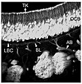
Description Figure legend provided in original article (CC-by): "This image shows a vertical projection of a stack of confocal images taken from a transgenic mouse, in which green fluorescent protein (GFP) is expressed in all ciliated olfactory sensory neurons. GFP brightly labels the olfactory neuronal cell bodies (OCB), their apical dendrites (AD), and terminal knobs (TK). Staining does not extend into the sensory cilia, which remain invisible in this preparation. Basally to the olfactory neuronal cell bodies is the unstained layer of basal stem cells (LBC), from which degenerating neurons are constantly regenerated. The olfactory axons grow in bundles (AB) through the basal lamina (BL), and then fasciculate to form the tracts of the olfactory nerve, which projects into the brain. (Rebecca Elsaesser and Jacques Paysan, unpublished)."
Date Prepared for Commons on 2007-12-18.
Source Elsaesser, Rebecca; Jacques Paysan (2007). "The sense of smell, its signalling pathways, and the dichotomy of cilia and microvilli in olfactory sensory cells". BMC Neuroscience 8 (Suppl 3): S1. DOI:10.1186/1471-2202-8-S3-S1. ISSN 1471-2202. Retrieved on 2007-12-18.
Author Prepared by User:OldakQuill from a CC-by figure in a journal article by Rebecca Elsaesser and Jacques Paysan (see source).
Permission
(Reusing this file)
w:en:Creative Commons
attribution
This file is licensed under the Creative Commons Attribution 2.0 Generic license.
You are free:
  • to share – to copy, distribute and transmit the work
  • to remix – to adapt the work
Under the following conditions:
  • attribution – You must give appropriate credit, provide a link to the license, and indicate if changes were made. You may do so in any reasonable manner, but not in any way that suggests the licensor endorses you or your use.

Captions

Add a one-line explanation of what this file represents

Items portrayed in this file

depicts

image/jpeg

File history

Click on a date/time to view the file as it appeared at that time.

Date/TimeThumbnailDimensionsUserComment
current22:57, 18 December 2007Thumbnail for version as of 22:57, 18 December 2007590 × 600 (178 KB)OldakQuill{{Information |Description=Figure legend provided in original article (CC-by): "This image shows a vertical projection of a stack of confocal images taken from a transgenic mouse, in which green fluorescent protein (GFP) is expressed in all ciliated olfac

Global file usage

The following other wikis use this file:

Metadata