File:Low-pressure-hyperbaric-oxygen-therapy-and-SPECT-brain-imaging-in-the-treatment-of-blast-induced-1757-1626-0002-0000006538-S1.ogv
Jump to navigation
Jump to search
Size of this JPG preview of this OGG file: 800 × 600 pixels. Other resolutions: 320 × 240 pixels | 640 × 480 pixels | 1,024 × 768 pixels.
Original file (Ogg Theora video file, length 14 s, 1,024 × 768 pixels, 634 kbps, file size: 1.08 MB)
| This free media file is from Wikimedia Commons. Its description page is included below. |
Summary
| Description |
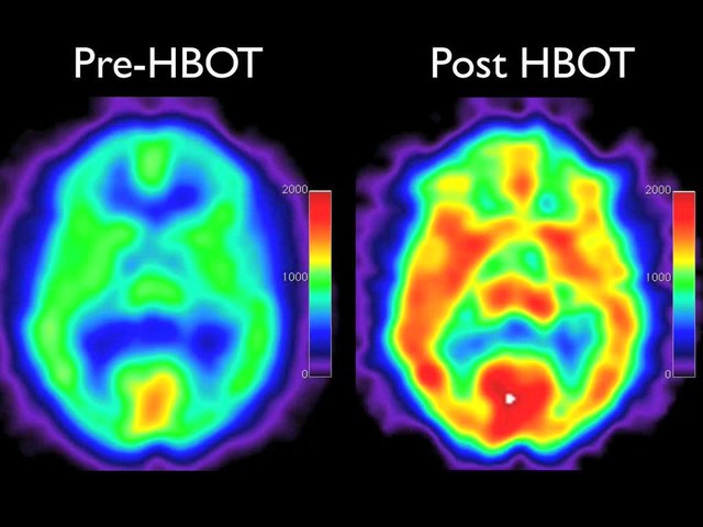
English: Side by side Pre and Post HBOT processed transverse SPECT brain blood flow images. Pre-HBOT scan is on the left and post-HBOT on the right. Click on either image to initialize movie. Images were obtained on a Picker Prism 3000 triple-head gamma camera. Both scans were processed by technologist PJT: 25 mCi of ECD was prepared with the standard manufacturer's kit and injected in a peripheral vein in a low noise low light area while the patient was quiet and motionless. One hour after injection acquisition proceeded with a 360 degree rotation and 40 stops, 20 seconds/stop on a 128 x 128 matrix, using low energy high resolution fan beam collimators. Motion correction was used for minor movement. Raw data was processed by transverse reconstruction using 360 degree filtered back projection and a ramp filter, followed by a LoPass filter, order 2.2. Cutoff was taken at the intersection of the best fit LoPass filter and noise on the power spectrum graph. Per file attenuation correction and best fit ellipse were applied. Images were oblique reformatted with slice thickness at 4 mm (2 pixels), aligned, and off-center zoom applied (20 cm2 area). Images were presented in all 3 orthogonal planes. Transverse processed images were analyzed with Osirix Open-source software (version 3.3.2) and windowed at a level of 1000 with a window width of 2000. They were subsequently rendered in QuickTime movie format starting from vertex and proceeding through the base of the brain. Images are in standard SPECT format and orientation. Color map is red, yellow, green, blue, and violet from highest brain blood flow to lowest. Note the marked generalized increase in perfusion on the post-HBOT scan |
||
| Date | |||
| Source | Harch P, Fogarty E, Staab P, Van Meter K (2009). "Low pressure hyperbaric oxygen therapy and SPECT brain imaging in the treatment of blast-induced chronic traumatic brain injury (post-concussion syndrome) and post traumatic stress disorder: a case report". Cases Journal. DOI:10.1186/1757-1626-0002-0000006538. PMC: 2740054. | ||
| Author | Harch P, Fogarty E, Staab P, Van Meter K | ||
| Permission (Reusing this file) |
This file is licensed under the Creative Commons Attribution 3.0 Unported license.
|
||
| Provenance |
|
File history
Click on a date/time to view the file as it appeared at that time.
| Date/Time | Thumbnail | Dimensions | User | Comment | |
|---|---|---|---|---|---|
| current | 14:38, 5 June 2013 | 14 s, 1,024 × 768 (1.08 MB) | Open Access Media Importer Bot | Automatically uploaded media file from Open Access source. Please report problems or suggestions here. |
File usage
The following 4 pages use this file: