File:Morbus Fabry kidney biopsy TEM 02.jpg
Jump to navigation
Jump to search

Size of this preview: 800 × 562 pixels. Other resolutions: 320 × 225 pixels | 640 × 449 pixels | 1,024 × 719 pixels | 1,181 × 829 pixels.
Original file (1,181 × 829 pixels, file size: 448 KB, MIME type: image/jpeg)
| This free media file is from Wikimedia Commons. Its description page is included below. |
| Description |
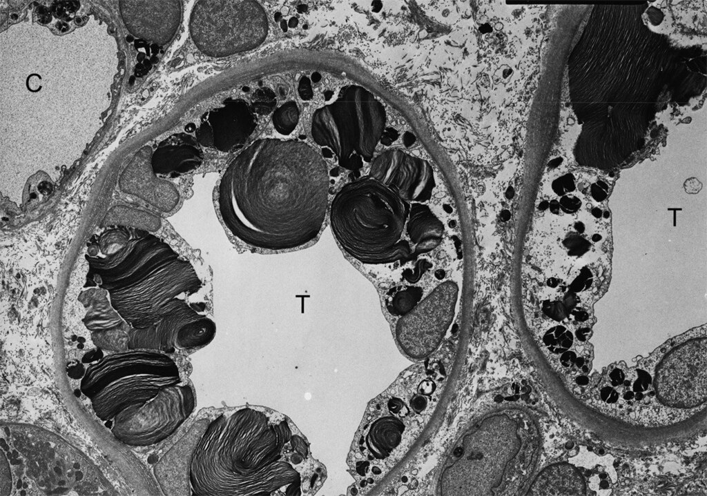
English: Kidney biopsy (electron microscopy): glycosphingolipid inclusions of various size and shape are seen in the cells of distal tubules of the kidney in Fabry disease.
Deutsch: Die Transmissionselektronenmikroskopische Aufnahme einer Gewebeprobe aus den Nieren eines Morbus-Fabry-Patienten zeigt die Inklusionen von Glycosphingolipiden unterschiedlicher Form und Größe in den Zellen des distalen Tubulus. |
| Date | article published: 22 November 2010 [1] |
| Source | D. P. Germain: Fabry disease. In: Orphanet journal of rare diseases Vol. 5, 2010, 30, PMID 21092187. PMC 300961. (Review) |
| Author | Juan M. POLITEI, Buenos Aires, Argentina; Dominique P Germain |
This file is licensed under the Creative Commons Attribution 2.0 Generic license.
- You are free:
- to share – to copy, distribute and transmit the work
- to remix – to adapt the work
- Under the following conditions:
- attribution – You must give appropriate credit, provide a link to the license, and indicate if changes were made. You may do so in any reasonable manner, but not in any way that suggests the licensor endorses you or your use.
File history
Click on a date/time to view the file as it appeared at that time.
| Date/Time | Thumbnail | Dimensions | User | Comment | |
|---|---|---|---|---|---|
| current | 19:18, 31 August 2011 | | 1,181 × 829 (448 KB) | Kuebi | {{Information |Description={{en|Kidney biopsy (electron microscopy): glycosphingolipid inclusions of various size and shape are seen in the cells of distal tubules of the kidney in Fabry disease. }} {{de|Die Transmissionselektronenmikroskopische Aufnahme |
File usage
The following 2 pages use this file:
Global file usage
The following other wikis use this file:
- Usage on de.wikipedia.org
- Usage on ru.wikipedia.org
- Usage on sr.wikipedia.org